Caesium »
PDB 2ov4-4ht3 »
3ump »
Caesium in PDB 3ump: Crystal Structure of the Phosphofructokinase-2 From Escherichia Coli in Complex with Cesium and Atp
Enzymatic activity of Crystal Structure of the Phosphofructokinase-2 From Escherichia Coli in Complex with Cesium and Atp
All present enzymatic activity of Crystal Structure of the Phosphofructokinase-2 From Escherichia Coli in Complex with Cesium and Atp:
2.7.1.11;
2.7.1.11;
Protein crystallography data
The structure of Crystal Structure of the Phosphofructokinase-2 From Escherichia Coli in Complex with Cesium and Atp, PDB code: 3ump
was solved by
H.M.Pereira,
A.Caniuguir,
M.Baez,
R.Cabrera,
R.C.Garratt,
J.Babul,
with X-Ray Crystallography technique. A brief refinement statistics is given in the table below:
| Resolution Low / High (Å) | 49.05 / 1.85 |
| Space group | P 2 2 21 |
| Cell size a, b, c (Å), α, β, γ (°) | 43.856, 88.914, 176.405, 90.00, 90.00, 90.00 |
| R / Rfree (%) | 17.2 / 20.9 |
Other elements in 3ump:
The structure of Crystal Structure of the Phosphofructokinase-2 From Escherichia Coli in Complex with Cesium and Atp also contains other interesting chemical elements:
| Magnesium | (Mg) | 4 atoms |
Caesium Binding Sites:
The binding sites of Caesium atom in the Crystal Structure of the Phosphofructokinase-2 From Escherichia Coli in Complex with Cesium and Atp
(pdb code 3ump). This binding sites where shown within
5.0 Angstroms radius around Caesium atom.
In total 2 binding sites of Caesium where determined in the Crystal Structure of the Phosphofructokinase-2 From Escherichia Coli in Complex with Cesium and Atp, PDB code: 3ump:
Jump to Caesium binding site number: 1; 2;
In total 2 binding sites of Caesium where determined in the Crystal Structure of the Phosphofructokinase-2 From Escherichia Coli in Complex with Cesium and Atp, PDB code: 3ump:
Jump to Caesium binding site number: 1; 2;
Caesium binding site 1 out of 2 in 3ump
Go back to
Caesium binding site 1 out
of 2 in the Crystal Structure of the Phosphofructokinase-2 From Escherichia Coli in Complex with Cesium and Atp
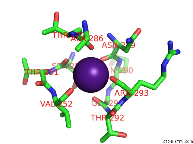
Mono view
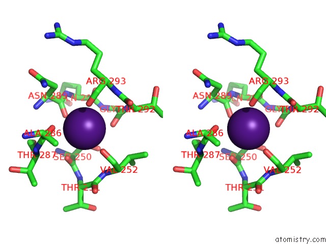
Stereo pair view
Mono view
Stereo pair view
A full contact list of Caesium with other atoms in the Cs binding
site number 1 of Crystal Structure of the Phosphofructokinase-2 From Escherichia Coli in Complex with Cesium and Atp within 5.0Å range:
|
Caesium binding site 2 out of 2 in 3ump
Go back to
Caesium binding site 2 out
of 2 in the Crystal Structure of the Phosphofructokinase-2 From Escherichia Coli in Complex with Cesium and Atp
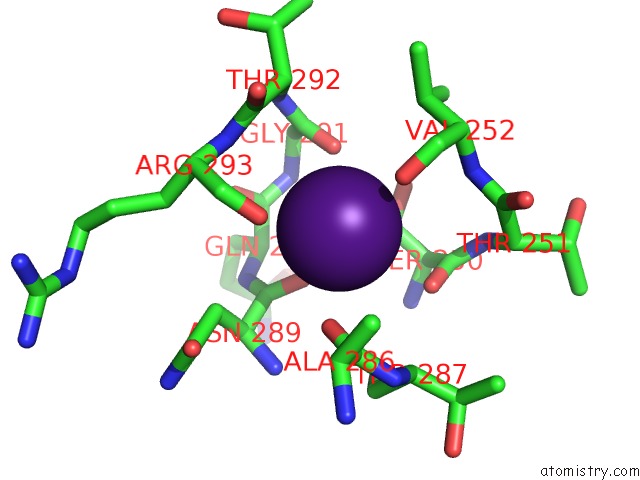
Mono view
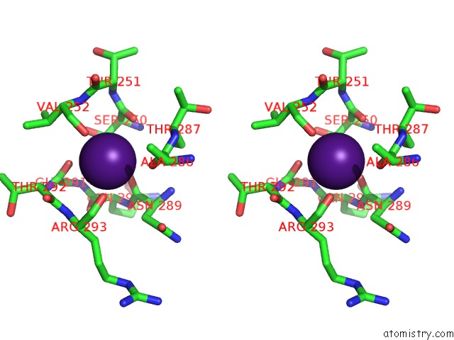
Stereo pair view
Mono view
Stereo pair view
A full contact list of Caesium with other atoms in the Cs binding
site number 2 of Crystal Structure of the Phosphofructokinase-2 From Escherichia Coli in Complex with Cesium and Atp within 5.0Å range:
|
Reference:
M.Baez,
R.Cabrera,
H.M.Pereira,
A.Blanco,
P.Villalobos,
C.A.Ramirez-Sarmiento,
A.Caniuguir,
V.Guixe,
R.C.Garratt,
J.Babul.
A Ribokinase Family Conserved Monovalent Cation Binding Site Enhances the Mgatp-Induced Inhibition in E. Coli Phosphofructokinase-2 Biophys.J. V. 105 185 2013.
ISSN: ISSN 0006-3495
PubMed: 23823238
DOI: 10.1016/J.BPJ.2013.05.028
Page generated: Tue Jul 30 20:24:17 2024
ISSN: ISSN 0006-3495
PubMed: 23823238
DOI: 10.1016/J.BPJ.2013.05.028
Last articles
Zn in 9MJ5Zn in 9HNW
Zn in 9G0L
Zn in 9FNE
Zn in 9DZN
Zn in 9E0I
Zn in 9D32
Zn in 9DAK
Zn in 8ZXC
Zn in 8ZUF