Caesium »
PDB 4hul-6dwe »
5dea »
Caesium in PDB 5dea: Crystal Structure of the Complex Between Human Fmrp Rgg Motif and G- Quadruplex Rna, Cesium Bound Form.
Protein crystallography data
The structure of Crystal Structure of the Complex Between Human Fmrp Rgg Motif and G- Quadruplex Rna, Cesium Bound Form., PDB code: 5dea
was solved by
N.Vasilyev,
A.Polonskaia,
J.C.Darnell,
R.B.Darnell,
D.J.Patel,
A.Serganov,
with X-Ray Crystallography technique. A brief refinement statistics is given in the table below:
| Resolution Low / High (Å) | 19.87 / 2.80 |
| Space group | P 21 21 2 |
| Cell size a, b, c (Å), α, β, γ (°) | 56.570, 129.870, 36.790, 90.00, 90.00, 90.00 |
| R / Rfree (%) | 20.2 / 22.6 |
Other elements in 5dea:
The structure of Crystal Structure of the Complex Between Human Fmrp Rgg Motif and G- Quadruplex Rna, Cesium Bound Form. also contains other interesting chemical elements:
| Potassium | (K) | 4 atoms |
Caesium Binding Sites:
The binding sites of Caesium atom in the Crystal Structure of the Complex Between Human Fmrp Rgg Motif and G- Quadruplex Rna, Cesium Bound Form.
(pdb code 5dea). This binding sites where shown within
5.0 Angstroms radius around Caesium atom.
In total 3 binding sites of Caesium where determined in the Crystal Structure of the Complex Between Human Fmrp Rgg Motif and G- Quadruplex Rna, Cesium Bound Form., PDB code: 5dea:
Jump to Caesium binding site number: 1; 2; 3;
In total 3 binding sites of Caesium where determined in the Crystal Structure of the Complex Between Human Fmrp Rgg Motif and G- Quadruplex Rna, Cesium Bound Form., PDB code: 5dea:
Jump to Caesium binding site number: 1; 2; 3;
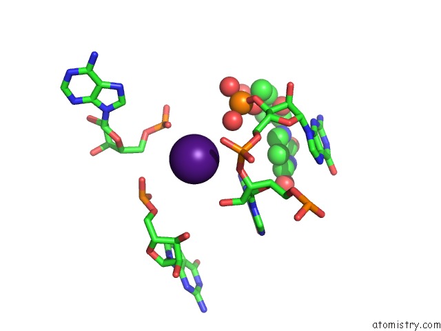
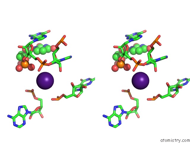
Caesium binding site 1 out of 3 in 5dea
Go back to
Caesium binding site 1 out
of 3 in the Crystal Structure of the Complex Between Human Fmrp Rgg Motif and G- Quadruplex Rna, Cesium Bound Form.
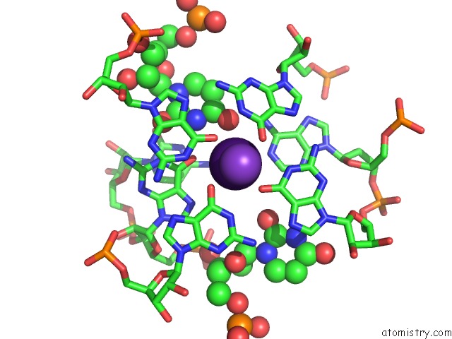
Mono view
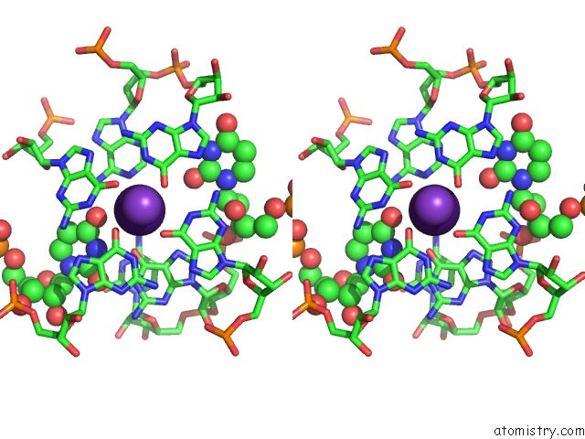
Stereo pair view
Mono view
Stereo pair view
A full contact list of Caesium with other atoms in the Cs binding
site number 1 of Crystal Structure of the Complex Between Human Fmrp Rgg Motif and G- Quadruplex Rna, Cesium Bound Form. within 5.0Å range:
|
Caesium binding site 2 out of 3 in 5dea
Go back to
Caesium binding site 2 out
of 3 in the Crystal Structure of the Complex Between Human Fmrp Rgg Motif and G- Quadruplex Rna, Cesium Bound Form.
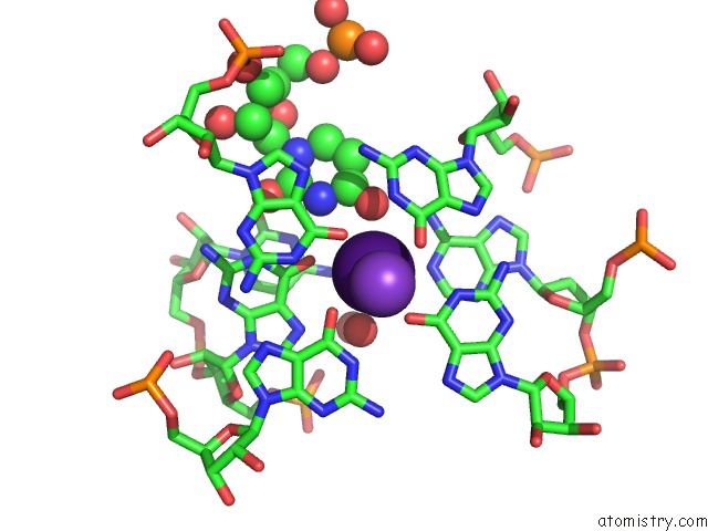
Mono view
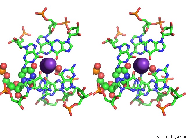
Stereo pair view
Mono view
Stereo pair view
A full contact list of Caesium with other atoms in the Cs binding
site number 2 of Crystal Structure of the Complex Between Human Fmrp Rgg Motif and G- Quadruplex Rna, Cesium Bound Form. within 5.0Å range:
|
Caesium binding site 3 out of 3 in 5dea
Go back to
Caesium binding site 3 out
of 3 in the Crystal Structure of the Complex Between Human Fmrp Rgg Motif and G- Quadruplex Rna, Cesium Bound Form.
Mono view
Stereo pair view
Mono view
Stereo pair view
A full contact list of Caesium with other atoms in the Cs binding
site number 3 of Crystal Structure of the Complex Between Human Fmrp Rgg Motif and G- Quadruplex Rna, Cesium Bound Form. within 5.0Å range:
|
Reference:
N.Vasilyev,
A.Polonskaia,
J.C.Darnell,
R.B.Darnell,
D.J.Patel,
A.Serganov.
Crystal Structure Reveals Specific Recognition of A G-Quadruplex Rna By A Beta-Turn in the Rgg Motif of Fmrp. Proc.Natl.Acad.Sci.Usa V. 112 E5391 2015.
ISSN: ESSN 1091-6490
PubMed: 26374839
DOI: 10.1073/PNAS.1515737112
Page generated: Sun Jul 13 22:49:51 2025
ISSN: ESSN 1091-6490
PubMed: 26374839
DOI: 10.1073/PNAS.1515737112
Last articles
Fe in 6MYPFe in 6MYO
Fe in 6MSN
Fe in 6MX5
Fe in 6MV0
Fe in 6MEV
Fe in 6MQ6
Fe in 6MQ1
Fe in 6MQ0
Fe in 6MPY