Caesium »
PDB 1av2-2j9x »
1kh8 »
Caesium in PDB 1kh8: Structure of A Cis-Proline (P114) to Glycine Variant of Ribonuclease A
Enzymatic activity of Structure of A Cis-Proline (P114) to Glycine Variant of Ribonuclease A
All present enzymatic activity of Structure of A Cis-Proline (P114) to Glycine Variant of Ribonuclease A:
3.1.27.5;
3.1.27.5;
Protein crystallography data
The structure of Structure of A Cis-Proline (P114) to Glycine Variant of Ribonuclease A, PDB code: 1kh8
was solved by
D.A.Schultz,
A.M.Friedman,
M.A.White,
R.O.Fox,
with X-Ray Crystallography technique. A brief refinement statistics is given in the table below:
| Resolution Low / High (Å) | 30.00 / 2.00 |
| Space group | P 43 21 2 |
| Cell size a, b, c (Å), α, β, γ (°) | 40.870, 40.870, 129.250, 90.00, 90.00, 90.00 |
| R / Rfree (%) | 19.5 / 21.9 |
Caesium Binding Sites:
The binding sites of Caesium atom in the Structure of A Cis-Proline (P114) to Glycine Variant of Ribonuclease A
(pdb code 1kh8). This binding sites where shown within
5.0 Angstroms radius around Caesium atom.
In total only one binding site of Caesium was determined in the Structure of A Cis-Proline (P114) to Glycine Variant of Ribonuclease A, PDB code: 1kh8:
In total only one binding site of Caesium was determined in the Structure of A Cis-Proline (P114) to Glycine Variant of Ribonuclease A, PDB code: 1kh8:
Caesium binding site 1 out of 1 in 1kh8
Go back to
Caesium binding site 1 out
of 1 in the Structure of A Cis-Proline (P114) to Glycine Variant of Ribonuclease A
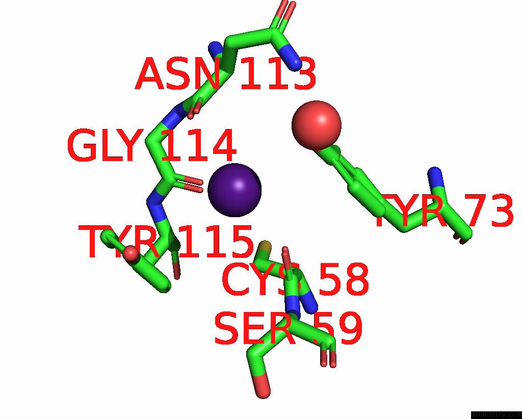
Mono view
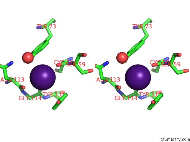
Stereo pair view
Mono view
Stereo pair view
A full contact list of Caesium with other atoms in the Cs binding
site number 1 of Structure of A Cis-Proline (P114) to Glycine Variant of Ribonuclease A within 5.0Å range:
|
Reference:
D.A.Schultz,
A.M.Friedman,
M.A.White,
R.O.Fox.
The Crystal Structure of the Cis-Proline to Glycine Variant (P114G) of Ribonuclease A. Protein Sci. V. 14 2862 2005.
ISSN: ISSN 0961-8368
PubMed: 16199662
DOI: 10.1110/PS.051610505
Page generated: Tue Jul 30 20:10:53 2024
ISSN: ISSN 0961-8368
PubMed: 16199662
DOI: 10.1110/PS.051610505
Last articles
Zn in 9MJ5Zn in 9HNW
Zn in 9G0L
Zn in 9FNE
Zn in 9DZN
Zn in 9E0I
Zn in 9D32
Zn in 9DAK
Zn in 8ZXC
Zn in 8ZUF